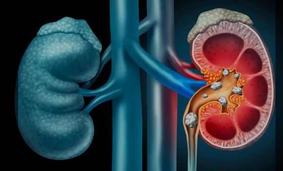
Kidney stones are hard deposits of minerals and salts that form inside the kidneys. They are a common urological condition that can cause severe pain, urinary problems, and, if untreated, serious complications such as infections or kidney damage. Managing kidney stones is essential to relieve symptoms, prevent recurrence, and protect long-term kidney health. The choice of management depends on the size, type, and location of the stone, as well as the patient’s symptoms and overall health.
The kidneys are two bean-shaped organs located on either side of the spine. Their primary role is to filter blood, remove waste products, and regulate electrolytes and fluid balance. Urine formed in the kidneys travels through the ureters to the bladder before being excreted. Kidney stones develop when minerals (such as calcium, oxalate, or uric acid) crystallize and clump together, sometimes obstructing urine flow. Stones can remain in the kidney or move into the ureter, causing intense pain known as renal colic.
Kidney stones form due to an imbalance in urine composition, often when urine becomes too concentrated. Common risk factors include:
Dehydration – Not drinking enough fluids increases urine concentration.
Diet – High intake of salt, animal protein, or foods rich in oxalate (e.g., spinach, nuts).
Medical conditions – Gout, obesity, inflammatory bowel disease, or urinary tract infections.
Family history – Genetics can increase susceptibility.
Certain medications – Some diuretics, calcium-based antacids, or antivirals may raise stone risk.
Metabolic abnormalities – Hypercalciuria (high calcium in urine) or hyperuricosuria (high uric acid).
Symptoms vary depending on stone size and location, but common signs include:
Severe, sharp pain in the back, side, or lower abdomen (renal colic).
Pain radiating to the groin or genital area.
Frequent or painful urination.
Blood in urine (hematuria).
Nausea and vomiting.
Fever and chills (if infection is present).
Difficulty passing urine if there is complete blockage.
Diagnosis involves a combination of clinical evaluation and imaging.
Medical history and physical exam – To identify risk factors and assess pain severity.
Urinalysis – Detects blood, crystals, or infection.
Blood tests – Checks kidney function, calcium, and uric acid levels.
Imaging studies:
Ultrasound – Non-invasive and safe, especially for pregnant women.
CT scan (non-contrast helical CT) – Gold standard for detecting stones.
X-ray (KUB) – Can detect some types of stones but less sensitive.
Management depends on stone size, location, composition, and patient condition.
Observation & hydration – Small stones (<5 mm) often pass naturally with increased fluid intake.
Pain management – NSAIDs or opioids may be prescribed.
Medical Expulsive Therapy (MET) – Medications like alpha-blockers (e.g., tamsulosin) can relax the ureter, facilitating stone passage.
Dietary changes – Reducing salt, oxalate-rich foods, and increasing water intake.
Extracorporeal Shock Wave Lithotripsy (ESWL) – Uses sound waves to break stones into smaller fragments that pass naturally.
Ureteroscopy with Laser Lithotripsy – A thin scope inserted into the ureter allows laser fragmentation of the stone.
Percutaneous Nephrolithotomy (PCNL) – Minimally invasive surgery for large stones (>2 cm), involving direct removal through a small incision in the back.
Open or laparoscopic surgery – Rarely needed, reserved for complex or recurrent cases.
ESWL: The patient lies on a water-filled cushion; targeted shock waves break the stone into pieces, typically performed as an outpatient procedure.
Ureteroscopy: A small scope is passed through the urethra and bladder into the ureter; the stone is fragmented using a laser and removed with a basket if needed.
PCNL: Under general anesthesia, a small incision is made in the back, and instruments are inserted directly into the kidney to remove large stones.
Recovery depends on the type of treatment:
After ESWL – Patients may pass stone fragments in urine for days to weeks; drinking plenty of fluids helps.
After ureteroscopy – A temporary stent may be placed to ensure urine flow; mild discomfort is common.
After PCNL – Hospital stay of 1–2 days is typical; patients may need a nephrostomy tube for drainage temporarily.
General recovery tips:
Stay hydrated to flush out stone fragments.
Take prescribed pain relief and antibiotics if needed.
Follow up with imaging to ensure stone clearance.
Adopt dietary modifications to reduce recurrence risk.
While most treatments are safe, potential risks include:
Infection or sepsis.
Bleeding or injury to urinary tract structures.
Residual or recurrent stones.
Stent-related discomfort (if placed after ureteroscopy).
Kidney damage (rare in modern procedures).
With timely treatment, prognosis is excellent. Small stones often pass spontaneously, while larger stones can be effectively treated with minimally invasive procedures. However, recurrence is common—around 30–50% within 5 years—so preventive measures (hydration, diet, lifestyle changes) are crucial.
Seek medical attention if you experience:
Severe flank or abdominal pain unrelieved by medication.
Blood in urine or difficulty urinating.
Fever, chills, or signs of infection.
Nausea and vomiting with dehydration.
Recurrence of kidney stone symptoms.
Kidney stones are a painful but manageable condition. Treatment may range from conservative approaches like hydration and medication to minimally invasive procedures such as ESWL, ureteroscopy, or PCNL. Long-term prevention, through lifestyle and dietary changes, plays a key role in reducing recurrence. If you suspect kidney stones, consult a healthcare professional for timely diagnosis and appropriate management to protect kidney health.
Chat With Me